
Anatomy Tips for PEBC EE
By Phartoonz
3 min read
Key takeaways
Anatomy tips for the PEBC Evaluating Exam: body movements, tissue types, tendon vs ligament vs cartilage, anatomical planes, joints and muscle groups.
Basic concepts & tips of anatomy should be covered in PEBC EE, according to the blue print for EE Physiology/ Functional Anatomy is included and should be revised.
Let’s go – EE anatomy tips:
Body Movements:
- abduction : moving a limb away in the frontal plane from the median plane of the body, spreading the fingers apart.
- adduction : opposite of abduction, movement of a limb toward the body mid-line.
- flexion : Bending of a part of the body i.e (decreases the angle of the joint and brings two bones closer together).
- extension : opposite of flexion; movement in the saggital plane that increases the angle of the joint or distance between two bones or parts of the body.
- dorsiflexion : lifting the foot so the superior surface approaches the shin, standing on the heels, moving upward.
- plantar flexion : pointing the toes.
- Pronation : Turning the hand so that the palm is down.
- Supination : Turning the hand so that the palm is up.
- Proximal: Closer to the point of attachment along the axis.
- Distal: Further from the point of attachment along the axis.
- Lateral view : from the side of the body.
- Medial view : from the middle of body.
- Superior: Up/above along the axis of upright posture (upwards in standard anatomical position). This is also called “Cranial” or sometimes “Cephalic”.
- Inferior: Down/below along the axis of upright posture. This is equivalent to “Caudal”.
- Anterior: towards the belly side. This is also known as “Ventral”
- Posterior: towards the back side. This is also known as “Dorsal”
Illustration By Video
Types of Tissues:
- Epithelial Tissue : Covers body and organ surfaces, lines body cavities, and form glands. Involved with protection, absorption, excretion, secretion, diffusion and filtration.
- Connective Tissue : binds , supports and protects body parts & stores energy and minerals.
- Muscle Tissue : Skeletal, cardiac and smooth muscle tissues. (Movement Functions)
- Nervous Tissue : Transmits nerve impulses and co-ordinates body activities.
Tendon vs Ligament vs Cartilage :
- Tendons attach the muscles to bones.
- Ligaments connect bones to bones at the joint.
- Cartilage is a substance which covers the ends of the bones.
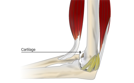
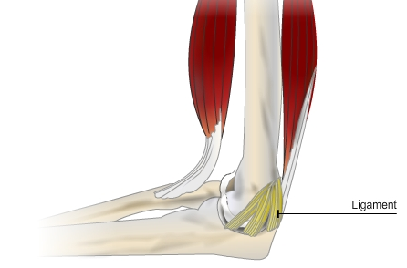
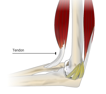
Source: bbc.co.uk
Anatomical planes : 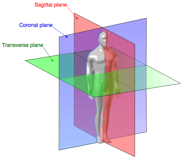
Coronal Plane (Frontal Plane) : A vertical plane running from side to side, it divides the body or any of its parts into anterior and posterior portions.
Sagittal Plane (Lateral Plane) : A vertical plane running from front to back, it divides the body or any of its parts into right and left sides.
Axial Plane (Transverse Plane) : A horizontal plane; divides the body or any of its parts into upper and lower parts.
Median plane : Sagittal plane through the midline of the body, it divides the body or any of its parts into right and left halves.
Body Positions :
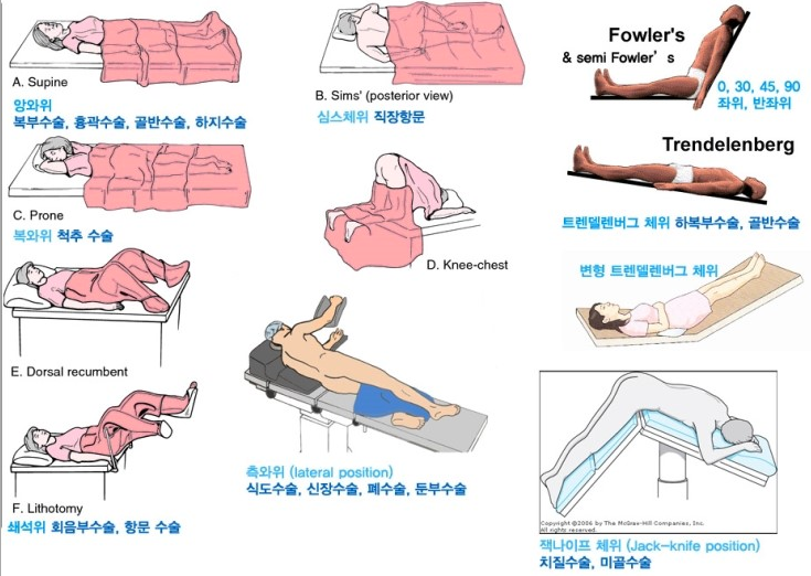
Covered before but I’m going to explain 2 important positions :
Fowler’s position : Categorized high, semi & low Fowler’s position based on sitting angle. it is seated position with back support ( Remember it by hospital bed and you control it by angle).
Trendelenburg position : Head is lowered and feet are elevated.
Body Joints:
|
Joint Name |
Bones of the Joint |
Type of Movement |
| Shoulder | Humerus, scapula and clavicle | Flexion, extension, horizontal flexion, horizontal extension, abduction, adduction, rotation, circumduction, elevation, depression, protraction and retraction |
| Elbow | Humerus, radius and ulna | Flexion and extension |
| Wrist | Radius, ulna and carpals | Flexion, extension, abduction, adduction and circumduction |
| Sacroiliac | Sacrum and ilium | Very limited range due to strong ligaments |
| Hip
(ball and socket synovial joint) |
Femur and pelvis | Flexion, extension, horizontal flexion, horizontal extension, abduction, adduction, rotation and circumduction |
| Knee | Femur, patella, tibia and fibula | Flexion and Extension |
| Ankle | Tibia, fibula and tarsals | Plantar flexion, dorsiflexion, inversion, eversion, supination and pronation |
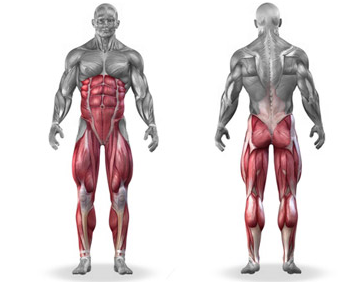
Major Muscle Groups:
You have to know the location of each muscle & direction either posterior or anterior.
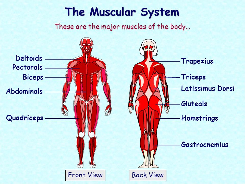
Tips to be continues with quizzes!
Keep reading
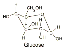
Quick important tips about Carbohydrates (Biochemistry)
Updated: 19/4/2017 So related to biochemistry we will review carbohydrates for the EE, we will share the most important tips and notes related to the PEBC pharmacits evaluating exam! Quick notes: Carbohydrates are the main sources of energy in the body. The liver has the major responsibility for maintaining blood glucose levels. The brain oxidizes…
Apr 18, 2017

PEBC Evaluation exam (EE)
PEBC Pharmacists Evaluating Exam In collaboration from pharmacists who have attended the Evaluation exam (EE) , we are going to start a new category of phartoonz.com to address latest questions, tips and study resources. (PEBC Forum) So for those who don’t know the Canadian Evaluation exam for pharmacists, it is the first step to get…
Apr 18, 2017
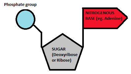
Macro-molecules nucleic acids structure
Following up wih PEBC notes and their evaluating exam structure, we will review a quick important notes as well as tips that you might be asked about during the exam. We will talk about the structure and important tips about the macro-molecules: Nucleic Acids The basic units of nucleic acids are nucleotides; each nucleotide contains…
Apr 22, 2017
Interactive Eye Anatomy
[kml_flashembed publishmethod=”static” fversion=”8.0.0″ movie=”http://phartoonz.com/phartoonZuploads/Eye-Anatomy.swf” width=”800″ height=”600″ targetclass=”flashmovie”] [/kml_flashembed] The most popular questions ( FAQs ) about the eyes from Google : What part of your eye is the iris? The colored part of the eye is the iris. It controls light levels inside the eye . The round opening in the center of the iris…
Oct 21, 2015